
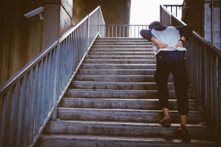
Athletes take heed – the Iliopsoas lurks as a major hidden root cause of pain for the lower back, hip and thigh/leg. Dr. Janet Travell, the mother of myofasical pain and trigger point medicine, named this muscle appropriately as the “Hidden Prankster”! Track and Field athletes, cyclists, triathletes, dancers and tennis players are all susceptible to this Iliopsoas Syndrome due to the repetitive hip flexion movements.

This muscle is actually comprised of two muscles: the psoas muscle and the iliacus muscle. They are synergistic in function in that they are primary hip flexors. However, due to its attachment along the spine, the iliopsoas plays a major role in maintaining upright posture. In addition, it also assists in lumbar spine movement and influences its ideal lordosis (curve). Both muscles coalesce and insert into the lesser trochanter of the femur at the inner, upper hip in the inguinal region (groin).
The potential regions of pain due to Iliopsoas dysfunction are the following:
Due to the depth of this muscle tissue, it may require some active rest and avoidance for a few of weeks to allow for the acute inflammation to subside (R.I.C.E.S.). The Iliopsoas is buried under the abdominal wall muscles. Its attachment is also difficult to access due to the upper inner femur insertion. However, soft tissue release, muscle energy and PNF (Proprioceptive Neuromuscular Facilitation) stretching will facilitate healing of this injury.
Frequently and all too often, doctors misdiagnose Iliopsoas Syndrome due to its pain referral patterns to the hip, lower back and thigh. As a result, a detailed examination via Applied Kinesiology and Biomechanics, as well as a detailed history of occurrence is necessary to pin this issue down. If this injury is at the insertion at the upper inner femur, cross fiber massage will be highly efficacious for resolution. This is done to re-align the tendon fiber attachment, thus avoiding a weak scar tissue adhesion.
The biomechanics must be addressed for the athlete. In addition, a joint clearing chiropractic session is frequently necessary to remove any distortion patterns in the kinetic chain from feet through pelvis! Again, pelvic distortion, joint restrictions, foot pronation and hip and knee imbalances will all perpetuate this pathology and must be addressed with manual therapy, possibly orthotics and of course facilitative stretching and eventually core stabilization exercises. For recalcitrant cases, MRI should be performed to isolate the degree of the injury and appropriate measures taken based on findings.
As with any inflammation and soft tissue injury support the healing process with proteolytic enzymes (Wobenzyme/Protrypsin), Collagen/Connective tissue nutrients (Ligaplex/Collagenics), Fish Oil and anti-inflammatory herbs such as boswellia, turmeric and ginger.
Finally, to prevent the dreaded re-occurrence, maintain proper alignment, joint support and nutrition an always train with excellent technique and warm-up/cool down procedures or get a gait analysis for detection of faults and contributing factors.
Image: Beth ohara
Monday – Friday: 5:30 am – 8:30 pm
Saturday – Sunday: 7:00 am – 3:00 pm
1834 George Avenue
Annapolis, MD 21401
Tel: (410) 224-7220
info@evolutionsannapolis.com